

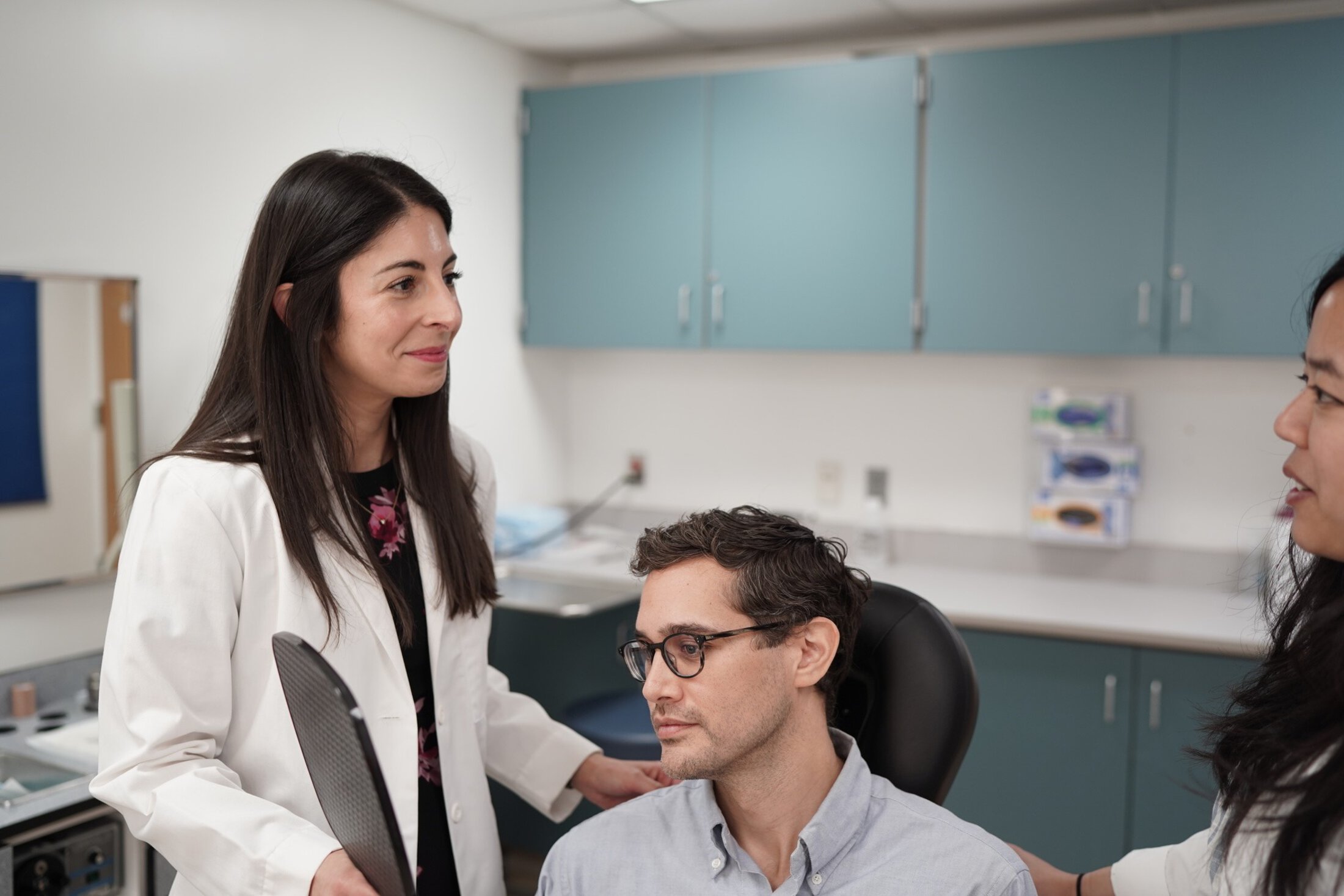
Michelle Hwang, M.D.
Specialties
- ENT - Facial Plastic & Reconstructive Surgery
- ENT - Ear, Nose & Throat
Locations (1)
- Charleston, SC
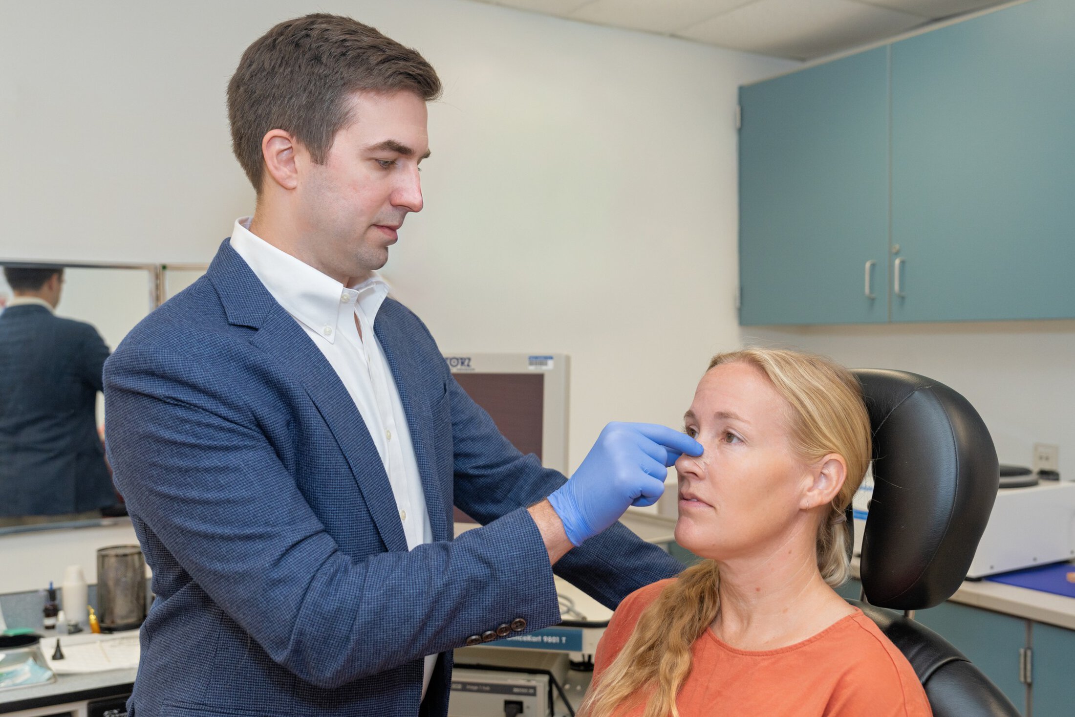
The MUSC Health Facial Nerve Center is South Carolina's only comprehensive center dedicated to the treatment of facial paralysis
Facial paralysis occurs when the muscles of the face become partially or completely weak. Our facial muscles play an essential role in expression, appearance, communication, and daily functions, such as blinking, nasal breathing, speaking, and eating. Facial paralysis can affect people of any age and may develop for many different reasons.
Identifying the underlying cause is a critical step. The recommended treatment approach changes depending on the cause of paralysis. Management is highly individualized and often involves a detailed conversation with your care team to review the risks, benefits, and goals of each treatment option.
The facial nerve is the seventh cranial nerve (CN VII). It begins in the brain and travels through a small canal in the skull alongside the nerve that gives hearing and balance. From there, it passes behind the eardrum and continues through the mastoid bone behind the ear. The nerve then exits the skull under the ear and enters the face passing through the parotid gland (the large saliva gland in the cheek). The nerve then divides into multiple branches that travel under the skin across the face and connect to the muscles responsible for facial movement.
The face contains more than 40 muscles that work together to create expressions. For these muscles to move normally, communication between the brain, the facial nerve, and the muscles must remain intact. Think of it like a lamp: it only works when the outlet, cord, and bulb are all functioning correctly. A problem anywhere along this pathway can lead to facial weakness or paralysis.
Facial paralysis can happen for many different reasons. In general, these causes fall into three main groups: congenital (present at birth), acute (developing suddenly), and chronic (developing slowly over time).
Bell’s palsy is the most common diagnosis of facial paralysis . Bell’s palsy is considered a diagnosis of exclusion, meaning all other causes should be reasonably ruled out. The weakness comes on relatively quickly and usually affects one entire side of the face. Some people with Bell’s palsy can move their facial muscles a little bit while others cannot move their muscles at all. With paralysis there may be associated pain in the ear, sensitivity to loud noises, changes in taste, numbness on the same side of the face, or changes in tear production. In addition to lack of facial expression, people with Bell’s palsy can also have difficulty with speech, eating, eye closure and breathing through their nose.
Sudden facial weakness can be alarming, and many people initially worry they are having a stroke. Anyone suffering from acute facial paralysis should seek immediate medical attention to identify the cause of the paralysis and begin treatment. The exact cause of paralysis in Bell’s palsy is not known, but it is thought to be related to swelling of the facial nerve due to a viral infection as it passes through a very narrow opening in the bone on its way from the brain to the facial muscles. The most effective treatment for Bell’s palsy is a course of steroids to counteract the swelling and inflammation around the nerve. There may also be a benefit from antiviral medication in addition to steroids. It is very important that individuals who cannot fully close their eye start using eye lubrication treatment to prevent dryness from damaging the eye.
For most people the facial movement begins to return within a few weeks of paralysis, but a minority of people do not see return of function begin for several months. Fortunately, a large majority of people will return to normal or near normal facial function following Bell’s palsy, but up to 30% of people will not full recover. Once facial movement begins to return, personalized facial massage and retraining exercises with a physical therapist may help improve outcomes. Incomplete recovery from facial paralysis can include decreased movements, abnormal tightness, involuntary movements (synkinesis), or spasms. There may be nonsurgical and surgical treatment options for those patients who do not fully recover.
Understanding the cause of facial paralysis is the first step in deciding the best treatment. In some cases, the facial movement may improve on its own with time. In other situations, weakness may not improve on its own and treatment may be time sensitive. Treatment options may include medications, physical therapy, minimally invasive injections, or surgery. Treatment is always individualized to the patient’s needs and the specifics of their condition.
Facial paralysis can impact many aspects of a person’s daily life, from appearance and facial expression to speech, eating, and emotional well-being. That is why treatment goals focus on restoring as much facial symmetry and function as possible. The goal is to help patients regain confidence and live a normal life without the physical or social challenges that can come with facial paralysis.
Facial synkinesis is an involuntary movement that happens when you are trying to make a voluntary facial expression, such as smiling. It can develop as the facial nerve heals after an injury.
As the nerve regenerates, some fibers may reconnect to the wrong muscles. When this happens, a signal meant for one muscle is unintentionally sent to others. For example, when you smile, your eye on the affected side may close. Or when you gently close your eyes, the corner of your mouth may lift.
Think of it like crossed telephone wires – one message is sent, but multiple lines pick it up. There are many treatment options for synkinesis, including facial rehabilitation and chemodenervation injections.
For some patients, medical therapy and physical rehabilitation may not provide enough improvement, and surgery may be recommended to help restore facial symmetry and function. Surgical approaches are often tailored to specific areas of the face and can be classified as either static or dynamic reconstruction. Static reconstruction involves repositioning or supporting facial features to improve appearance and function, but it does not restore movement. Dynamic reconstruction aims to restore movement to the affected areas. In certain cases, depending on the type of location of paralysis, static reconstruction may be the best option to achieve improved facial appearance and function.
Facial reanimation refers to surgical procedures that help restore movement and symmetry to your face.
These procedures not only improve resting facial posture, but also aim to improve function. For example, improved ability to smile, speak, eat, and improved ability to close your eye. Our goal is to help patients feel more like themselves again. A smile plays a major role in how we connect with others, restoring a natural looking smile is often the central focus of our treatment.
A smile is a key part of how we show emotion, communicate socially, and express our identity. When someone cannot smile or has the appearance of an uneven smile, it can impact confidence, emotional well-being, and daily interactions. When someone experiences facial paralysis, their muscle movements become uncoordinated and their function, in addition to lack of smile, becomes greatly impaired. Patients experience challenges with eating and drinking, closing the eye fully, and lack of resting facial symmetry. Our team provides a customized treatment plan to help maximally restore both function and appearance in the safest and most effective way possible.
These procedures are often used in combination to produce the best results. All nerve transfer procedures require at least six months for nerve healing and patient retraining to optimize results.
Selective Neurectomy
Hypoglossal to Facial (12 to 7) Nerve Transfer
Temporalis Tendon Transfer
Fascia Lata Facial Suspension
Asymmetric Facial Reconstruction or Facelift
Platysma Myectomy
Eyebrow Lift
Lip Lift
Depressor Anguli Oris (DAO) Myectomy
Masseteric to Facial (5 to 7) Nerve Transfer
Cross-Facial Nerve Grafts
Temporalis Tendon Transfer
Gracilis Free Muscle Transfer
Upper Eyelid Weight Placement
Lateral Tarsal Strip / lower eyelid tightening
Bipedicled Orbicularis Oculi Myocutaneous (BOOM) Flap
Modified Hughes Flap for lower eyelid tightening
Fascia Lata Facial Suspension
Facial paralysis can cause weakness to the muscle around the nose, leading to blocked nasal airflow. Tensor fascia lata is strong, natural tissue from your outer thigh. In this procedure, your surgeon carefully removes a strip of this tissue from your thigh and uses it to support or suspend certain droopy areas of the face such as the nose, corner of your mouth, cheek, or lower eyelid. This provides immediate improvement in facial symmetry at rest and helps with functions like breathing through your nose, eating, drinking, and speaking. This procedure does not provide additional ability to create volitional movements.
Septorhinoplasty
Facial paralysis can cause weakness of the muscles around the nose leading to blocked nasal airflow. A septorhinoplasty straightens the nasal septum and strengthened other parts of your nose with cartilage grafts to improve breathing through your nose. This is a comprehensive approach that addresses both the structural and functional problems that contribute to breathing difficulties in facial paralysis patients.
For many patients, facial paralysis can be meaningfully improved without surgery. Non-surgical treatment is tailored to the cause and stage of paralysis and generally falls into three categories: medical management (such as steroids or antivirals, most effective when started early), injectable treatments (botulinum toxin and facial filler), and rehabilitative therapy (physical therapy, speech therapy, and biobehavioral medicine) — together helping to reduce muscle tightness, restore symmetry, and support long-term function. In many cases, non-surgical treatment provides significant improvement, though some patients may go on to consider surgical options if further improvement is needed.

In some cases of facial paralysis, starting treatment early may help improve recovery. These treatments depend on the cause of the facial paralysis and can include medications such as antibiotics, antivirals, and steroids. For patients diagnosed with Bell’s Palsy, research shows that treatment with steroids and antiviral medication within 72 hours of onset likely improves recovery time and chances of full recovery. However, not all causes of facial paralysis benefit from medication, which is why it is important to undergo evaluation as soon as paralysis develops.
There are two main types of in-office injections used to help people with facial paralysis; these are chemodenervation with botulinum toxin and injectable filler. Both work in different ways and may be used together to help optimize facial function.
Chemodenervation with Botulinum Toxin
Botulinum tox is a well-studied and overall safe medication that has been used for years for many cosmetic and medical concerns. This medication works by temporarily weakening the muscle it is injected into. For the treatment of facial paralysis, very small doses are injected into muscles in the face primarily to help decrease excess muscle tightness, improve facial symmetry, and decrease other unwanted facial movements (synkinesis). By selectively weakening muscles that are overactive the function and balance of the face can be improved.
The injections are easily performed in the office with only minor discomfort. The results take one to two weeks to reach full effect and wear off after approximately three months. Repeat treatments are very effective and safe for years if needed. Patients have the best results when they combine botulinum toxin injections with facial retraining exercises. Generally, we are successful at getting botulinum toxin injections covered by your insurance plan.
For someone seeking a more permanent solution, a selective neurectomy may be an option.
Facial Filler
Injectable filler comes in many forms. It helps restore volume to various parts of the face and has been used for years in both cosmetic and reconstructive applications. Filler can be used in patients with facial paralysis to help restore facial symmetry and decrease liquid that may spill from the corner of the mouth.
Facial filler treatment is easily performed in the office with topical anesthesia and minimal discomfort. The effects of the injections are immediate, and most fillers will last between six months to two years.
Please note that when fillers are used to improve facial symmetry in patients with facial paralysis, they are not covered by insurance and are considered an out-of-pocket expense.
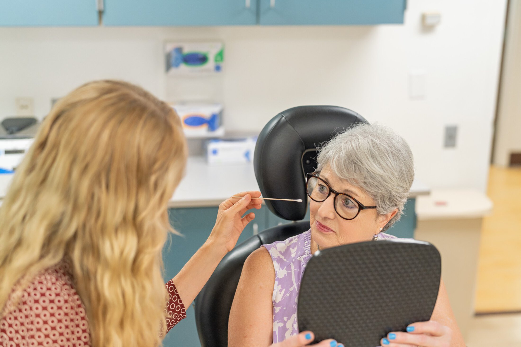
Physical therapy plays an important role in the evaluation and treatment of facial paralysis. Facial retraining is a distinct area of practice within physical therapy, guided by clinicians with focused training in facial nerve rehabilitation to support improved facial movement and function. This approach relies on active participation and consistent practice to promote meaningful, long-term improvement.
Treatment focuses on improving facial movement, coordination, and comfort while addressing symptoms such as weakness, asymmetry, muscle tightness, involuntary movements (synkinesis), pain, fatigue, difficulty with facial expressions, speech, eating, and eye protection. Sessions may include gentle facial massage and relaxation techniques to reduce muscle tension, along with targeted exercises designed to restore movement and symmetry.
Because normal facial expressions are small and subtle, retraining emphasizes precise, controlled movement to promote healthy nerve-to-muscle communication and reduce maladaptive movement patterns. Visual feedback, such as mirror therapy, is often used to help patients recognize, refine, and balance facial movements on both sides of the face.
Education and self-management strategies are an essential part of therapy, empowering patients to continue progress outside of the clinic and throughout recovery. By combining evidence-based techniques with individualized care, physical therapy supports both functional recovery and quality of life, helping patients regain confidence, comfort, and control in everyday facial movements.
Facial paralysis affects the ability to move the lips and cheeks which have an impact on an individual’s ability to speak and eat. Assessment by a speech and language pathologist can identify specific difficulties for each person and may offer compensation strategies to help with these important functions. Assessments can also be helpful to follow an individual after treatment or during recovery to track improvement.
Biobehavioral medicine recognizes that recovery from facial paralysis involves more than nerve and muscle healing alone. It focuses on the connection between biological factors (such as nerve injury, muscle function, and healing), psychological factors (including emotional health, stress, and self-image), and behavioral factors (movement patterns, habits, and social interaction).
Facial paralysis can affect how a person eats, speaks, expresses emotion, and connects with others. These changes may also influence confidence, mental well-being, and daily behavior. Stress, anxiety, or altered movement patterns can, in turn, impact muscle tension, coordination, and overall recovery.
By incorporating a biobehavioral therapy approach into our treatment model, our care addresses both physical function and quality of life. Treatment may include education on stress and muscle awareness and strategies that support emotional resilience and long-term recovery. This comprehensive model allows us to treat the whole person, not just the facial nerve injury - resulting in more effective, compassionate, and sustainable care.
It is common that patients with facial paralysis also experience balance problems. MUSC’s dedicated Balance Center provides specialized evaluation and therapy to address dizziness, imbalance, and difficulty with coordination.
Our team works in a multidisciplinary manner, collaborating closely with neurology, otolaryngology, neurosurgery, and rehabilitation specialists to create a personalized treatment plan. The goal of balance therapy is to improve stability, reduce dizziness, and help you feel confident and safe in your daily activities.